Home

ținut muntos recrut livrare la domiciliu cell electron microscope întuneric Amazon Jungle Masa de pranz

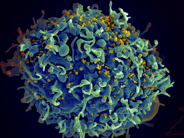
Multimedia Gallery - Scanning electron micrograph of HIV particles  infecting a human T cell. | NSF - National Science Foundation
Multimedia Gallery - Scanning electron micrograph of HIV particles infecting a human T cell. | NSF - National Science Foundation

A Photograph Of Mitochondria Showing All The Cells In It Stock Photo -  Download Image Now - iStock
A Photograph Of Mitochondria Showing All The Cells In It Stock Photo - Download Image Now - iStock

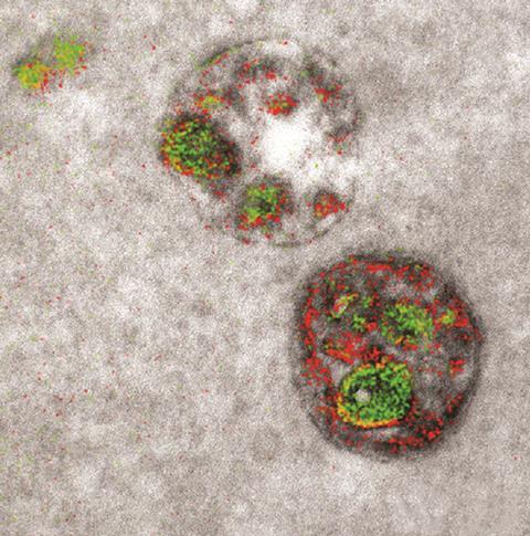
Transmission Electron Microscopy of Human Pluripotent Stem Cell Spheres...  | Download Scientific Diagram
Transmission Electron Microscopy of Human Pluripotent Stem Cell Spheres... | Download Scientific Diagram

Electron microscope captures cells in colour for the first time | Research  | Chemistry World
Electron microscope captures cells in colour for the first time | Research | Chemistry World

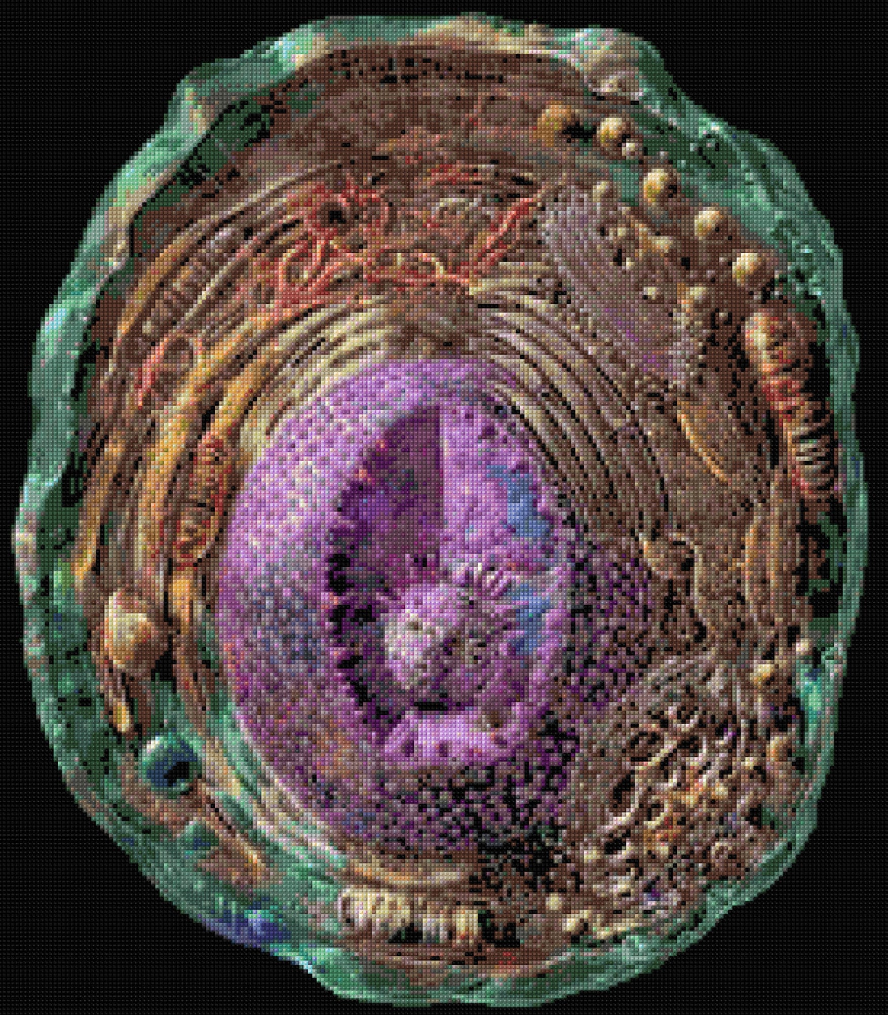
Microscopic Human Cell Electron Microscope Cross Stitch - Etsy Australia

2.3.3 Identify structures from electron micrographs of liver cells - YouTube

Electron Microscopy Goldfish Cell Stock Photo 756785854 | Shutterstock

File:Anatomy and physiology of animals animal cell electron microscope.jpg  - Wikimedia Commons
File:Anatomy and physiology of animals animal cell electron microscope.jpg - Wikimedia Commons

Electron microscopy of SARS-CoV-2: a challenging task - The Lancet

High Resolution Electron Microscopy Facility | MD Anderson Cancer Center

Tailoring staining protocols to electron microscopy workflows

Scanning Electron Microscopy Images of Cell Wall Structures after... |  Download Scientific Diagram
Scanning Electron Microscopy Images of Cell Wall Structures after... | Download Scientific Diagram

Transmission Electron Microscopy Reveals Distinct Macrophage- and Tick Cell-Specific  Morphological Stages of Ehrlichia chaffeensis | PLOS ONE
Transmission Electron Microscopy Reveals Distinct Macrophage- and Tick Cell-Specific Morphological Stages of Ehrlichia chaffeensis | PLOS ONE
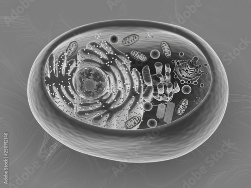
Animal cell, 3d rendering, Scanning Electron Microscope imitation texture  Stock Illustration | Adobe Stock
Animal cell, 3d rendering, Scanning Electron Microscope imitation texture Stock Illustration | Adobe Stock
What is a diagram of a plant and animal cell under an electron microscope?  - Quora
What is a diagram of a plant and animal cell under an electron microscope? - Quora

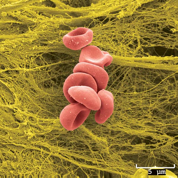
Scanning Electron Microscope Image of Blood Cells: Image Details - NCI  Visuals Online
Scanning Electron Microscope Image of Blood Cells: Image Details - NCI Visuals Online

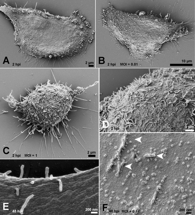
Ultrastructural analysis of SARS-CoV-2 interactions with the host cell via  high resolution scanning electron microscopy | Scientific Reports
Ultrastructural analysis of SARS-CoV-2 interactions with the host cell via high resolution scanning electron microscopy | Scientific Reports
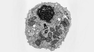
Electron microscopes - Cell structure - Edexcel - GCSE Combined Science  Revision - Edexcel - BBC Bitesize
Electron microscopes - Cell structure - Edexcel - GCSE Combined Science Revision - Edexcel - BBC Bitesize

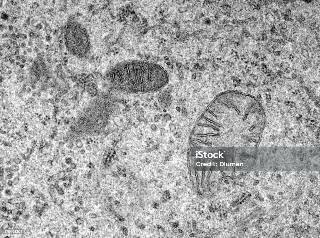
Scanning Electron Microscopy Images | Central Microscopy Research Facility

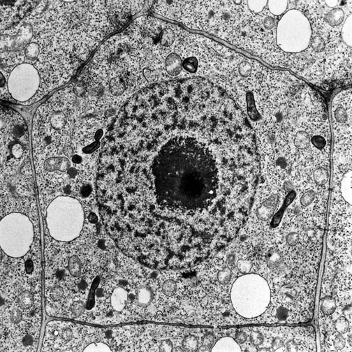
Electron Microscopy of Plant Cells (4.2) | Edexcel International AS Biology  Revision Notes 2018 | Save My Exams
Electron Microscopy of Plant Cells (4.2) | Edexcel International AS Biology Revision Notes 2018 | Save My Exams

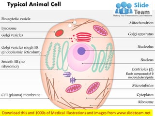
A typical animal cell (as seen in an electron microscope) Medical Ima…

AICE Biology Chapter 1: Animal Cell Electron Micrograph Labeling Diagram |  Quizlet
AICE Biology Chapter 1: Animal Cell Electron Micrograph Labeling Diagram | Quizlet

Electron microscopy (EM)

What is a diagram of a plant and animal cell under an electron microscope?  - Quora
What is a diagram of a plant and animal cell under an electron microscope? - Quora